The Role of Modern Imaging in Early Detection of Eye Diseases
The advancement of imaging technology has dramatically changed the way we identify and manage diseases of the eye. It will lead to improved diagnosis of eye diseases. For vision-threatening diseases, early detection will allow for a more intact vision at the end of the process of disease management. Some diseases may advance in the background without any clear indication. Early detection will ensure vision is preserved. OCT imaging eye, one of the new imaging technologies, is both accurate and non-invasive. It provides the ability for clinicians to obtain images of retinal cross-sections, which allows for the more comfortable observation of subtle changes.
Understanding OCT Imaging Eye Technology
Optical Coherence Tomography (OCT) is a non-invasive diagnostic imaging technology. It has transformed how near-infrared light waves can reveal detailed structures of the retina and optic nerve.
- OCT is similar to ultrasound imaging but uses light and not sound. It can take highly detailed images, with a precise focus on structures within the eye.
- With OCT imaging eye technology, images are represented in a three-dimensional visualisation. This shows the retina, optic nerve head, and surrounding ocular structures, highlighting the details of the retina. It has a resolution range of about 5 to 15 microns.
- The layer-by-layer resolution of the retina helps in portraying fine details about changes or abnormalities that may be missed with the naked eye or with grades of examination.
- The speed of the scan, the painless acquisition of information, and minimisation of patient involvement during the diagnostic process allow clinicians to conduct a proper examination without dilating the pupil in most cases.
- OCT imaging eye technology identifies early structural changes in the retina that can take place before any symptoms arise, leading to the availability of timely and clinically informed intervention.
The Critical Role of OCT Imaging Eye Worsley in Early Detection
Getting a head start with early detection of eye diseases is key to preventing irreversible vision loss, and OCT imaging eye Worsley clinics also provides access to this technology.
- OCT detects microstructure changes to the retina due to chronic disease. It is capable of assessing many eye-related diseases, such as glaucoma, age-related macular degeneration (AMD) and diabetic mellitus mediated retinopathy.
- These assessments give a quality baseline for your eye health, helping to describe any progress at future appointments.
- Provides quantitative measures relevant to intervention for eye conditions. This is also appropriate for evaluation of progression, such as retinal nerve fibre layer (RNFL) thickness and macular volume. It assists in the refinement of an individualised treatment plan based on the individual measures.
- It can improve patient prognosis by enabling interventions when damage is minimal or still reversible.
- By accessing OCT imaging eye, you benefit from not only an expert early diagnosis but also continued monitoring of your condition with potentially less likelihood of future complications.
Advantages of Using OCT Imaging Eye Worsley for Eye Health Management
When patients get their OCT imaging eye Worsley evaluations, they gain significant advantages, including:
- Non-invasive and Comfortable: No physical contact with the eye, pain-free, and, unlike most routine checks, typically doesn’t require pupil dilation.
- Highly Productive and Accurate Imaging: Provides objective and quantifiable measures that are standardised for staging and tracking measures of eye disease.
- Convenient and Timely Scans With Immediate Results: Scans take a few minutes, and the detailed report is reviewed immediately after the scan.
- Provides Active Monitoring of the Disease: Allows for longitudinal data collection with assessment of treatment response.
What to Expect During an OCT Imaging Eye Exam in Worsley
Knowing the process reduces apprehension and prepares individuals for the scan. Here’s how it takes place:
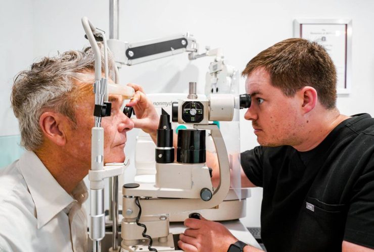
- Patients place their chin and forehead on the OCT machine’s chin rest.
- The OCT machine scans the retina and optic nerve using a low-intensity light beam in a non-invasive way.
- The scan is also not very long (5-10 minutes total).
- Generally, patients do not require pupil dilation, though a few may need it.
- The optometrist or ophthalmologist can view the scan right away and discuss the results by detailing what the scan means and what the next steps will be for the patient.
The OCT imaging eye service helps the community gain access to this advanced diagnosis of eye health for monitoring and easily documents incidents when problems arise.
Conclusion
OCT imaging eye technology represents a major advance in the early detection and management of many eye diseases. It allows clinicians to see beneath the surface with clarity that has never been possible before. Accessing an OCT imaging eye Worsley appointment is a pathway to early and timely diagnosis, custom treatment, and possibly a better vision prognosis. By choosing to use this imaging technology, we can catch eye ailments early before they become a major concern.
For expert care with cutting-edge OCT imaging eye worsley technology, you can be confident and choose only the qualified professionals at Robin Hall Opticians. Live your vision, with early detection being the first step!


